Skip to content

Lisa Klimas

I'm a 35 year old microbiologist and molecular biologist with systemic mastocytosis, Ehlers Danlos Syndrome, Postural Orthostatic Tachycardia Syndrome, Adrenal Insufficiency, and an assortment of other chronic health issues. My life is pretty much a blast.

DNA Methylation: How it works

DNA methylation is one of the ways your cells control which genes to express. It is an example of epigenetic modification. Epigenetics mechanisms like this do not change the DNA sequence, only the way the genes are expressed. Whether or not DNA methylation is heritable is not clear.

This is how DNA methylation works:

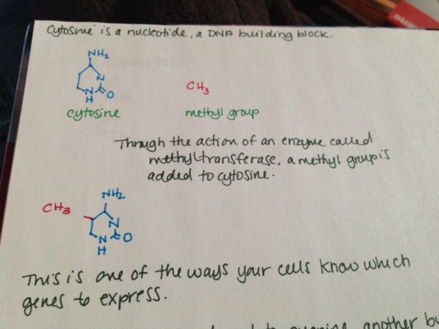
  • Cytosine is a nucleotide, a DNA building block.
  • Through the action of an enzyme called methyltransferase, a methyl group is added to cytosine.
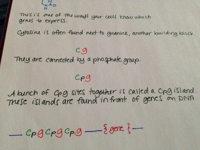
  • This is one of the ways your cells know which genes to express.
  • Cytosine is often found next to guanine, another building block. This is sometimes shown in literature as “CG.”
  • Cytosine and guanine are connected by a phosphate group. This is sometimes shown in literature as “CpG.”
  • A bunch of CpG sites together is called a CpG island. These islands are found in front of genes on DNA.
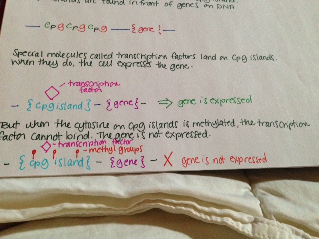
  • Special molecules called transcription factors land on CpG islands. When they do, the cell expresses the gene.
  • But when the cytosine on CpG islands is methylated, the transcription factor cannot bind. The gene is not expressed.

See pictures below.

Methylation is known to have an important role in cancer biology. Methylation of tumor suppressor genes causes the tumor suppressors not to be expressed, resulting in cancer.

Methylation 1Methylation 2Methylation 3

Lesser known mast cell mediators (Part 4)

Interleukin-1a (IL-1a) is largely responsible for inflammation, fever and sepsis. It activates TNF-a and the work very closely together. Their cofunctions include PGE2 synthesis, nitric oxide production, insulin resistance and IL-8 and chemokine production.

Interleukin-1b (IL-1b) has been implicated in several autoinflammatory syndromes. It is also important in cell proliferation, differentiation and apoptosis. Its induction of COX2 cytokine in the nervous system contributes to inflammatory pain hypersensitivity.

Interleukin-2 (IL-2) is crucial in prevention of autoimmune disease by regulating T cell differentiation. It is also thought to be involved in itchiness and psoriasis. IL-2 is used in the treatment of cancers.

Interleukin 3 (IL-3) drives the differentiation of multipotent hematopoietic stem cells into myeloid progenitor cells. If IL-7 is also present, they can work synergistically to trigger differentiation into lymphoid progenitor cells. IL-3 induces proliferation of all myeloid cells (including mast cells) along with other cytokines like IL-6. It supports growth and differentiation of T cells from bone marrow when an immune response is triggered.

Interleukin 4 (IL-4) changes naïve T cells to T helper cells, which secrete chemicals to drive actions of other immune cells. T helper cells then secrete additional IL-4 to perpetuate the cycle. IL-4 participates in the airway inflammation seen in allergic asthma.

Interleukin 5 (IL-5) encourages growth of B cells and antibody secretion as well as eosinophil activation. It is heavily involved in allergic diseases, particularly those in which eosinophils are notably increased. Mepolizumab is a monoclonal antibody against IL-5 that can reduce excessive eosinophils.

Interleukin 6 (IL-6) mediates fever and the acute phase inflammatory response. It is secreted to stimulate bone resorption and inhibitors of IL-6 are used to treat osteoporosis (including estrogen.) It inhibits TNF-a and IL-1. Unusually, it also has anti-inflammatory behaviors, particularly during exercise in the muscle.

Interleukin 9 (IL-9) increases cell proliferation and impedes apoptosis, cell death, of hematopoietic cells. It is particularly important in asthma and bronchial hyperresponsiveness.

Interleukin 10 (IL-10) is an anti-inflammatory molecule involved in regulating the JAK-STAT pathway. It counteracts many of the inflammatory effects of mast cells, often by interfering with production of substances like interferons and TNF-a.   Exercise increases levels of this molecule.

Interleukin 13 (IL-13) is critical in initiation of airway disease. It induces matrix metalloproteinases to act. IL-13 can also induce IgE release from B cells. It is effectively a link between allergic inflammatory cells and the non-immune cells they interact with. Excessive , IL-13 causes airway hyperresponsiveness, goblet cell metaplasia and oversecretion of mucus.


I think I might have mast cell disease: FAQ

What kinds of symptoms do mast cell patients have?

Mast cell disease can cause a variety of symptoms. Each person has their own unique constellation of complaints, and they can vary from day to day. Mast cell patients often have allergic type reactions to many things. They may have had anaphylaxis in the past, but that is not always the case.

What kind of doctor should I see if I think I have mast cell disease?

Due to the fact that mast cell disease can affect multiple body systems, it is managed by doctors of multiple disciplines. Immunologists, dermatologists, gastroenterologists and hematologists/ oncologists all may treat mast cell disease. It really depends who is familiar with mast cell disease in your areas. Immunologists are often the first stop for patients investigating mast cell disease.

Will any doctor know about mast cell disease?

No. Mast cell disease is uncommon. Many doctors are only aware of the types associated with pathologic rashes (cutaneous mastocytosis) or proliferation of mast cells in the bone marrow (systemic mastocytosis.)

How do I get determine if I have mast cell disease?

Labs for diagnosing mast cell disease include serum tryptase (a blood test), n-methylhistamine (24 hour urine test) and D2/F2a prostaglandin (24 hour urine test.) These tests are time sensitive for many patients and have special handling in most labs. Depending on these results, a bone marrow biopsy may be needed.

Can I have mast cell disease if my tryptase is normal?

Yes. 15% of patients with systemic mastocytosis have normal tryptase levels, and the majority of MCAS patients have normal tryptase levels.

How is mast cell disease treated?

Treatment generally focuses on the symptoms. The most common treatments include antihistamines, leukotriene inhibitors and mast cell stabilizers.

Will I feel better with treatment?

Most people feel better with treatment than without, but how much each person recovers is individual. Lifestyle modifications and medications can help many people live a full life.

Is mast cell disease curable?

No. Patients may have a remission from symptoms, but they will always have mast cell disease.

Symptoms of mast cell disease

The following is a generalized list of common symptoms associated with mast cell disease. It is not comprehensive and does not include laboratory or associated diagnoses.

General: fatigue, malaise (“being out of it”), weakness, severe unprovoked sweating, weight gain or loss

Skin: Rashes and lesions of any kind, itching, flushing, angioedema, stretch marks, dermatographism, poor wound healing, alopecia, abnormalities of finger or toenails

Eyes: Irritated eyes, red eyes, excessive tearing, dry eyes, difficulty focusing vision, lid tremor, sensitivity to lightness, including sunlight, general inflammation

Ears: Inflammation of the ear, “ear infections,” hearing loss, sensitivity to sound, ringing in the ears

Mouth/throat/sinuses: Generalized pain of several qualities, but often burning, ulceration, “canker” sores, swelling (angioedema), dental decay, abnormalities in taste, taste of metal, throat discomfort or irritation, need to clear throat frequently, post nasal drip, nose bleeds, irritation of sinuses, sinus congestion

Respiratory: laryngitis, bronchitis, pneumonitis (frequently confused with pneumonia), recurrent cough (usually dry), shortness of breath, wheezing

Cardiovascular: lightheadedness, weakness, dizziness, vertigo, fainting, high or low blood pressure, palpitations, rapid heartbeat, abnormalities in heart rhythm, chest pain, hemorrhoids, edema

Gastrointestinal: abdominal pain, diarrhea, constipation, difficulty swallowing, swelling of any part of the GI tract, nausea

Neurologic: Headache, migraine, “about to faint,” fainting, numbness, pins and needles, neuropathy, tics, tremors, seizures

Psychiatric: Anger, depression, PTSD, anxiety, memory difficulties, anxiety, panic disorders, insomnia, sleep disorders

The hallmark of mast cell disease is allergic-type reaction to a variety of stimuli. These can also occur to substances benign to most people, including scents, “hypoallergenic” materials, heat, sunlight and water. Some people experience anaphylactic reactions that require epinephrine.

References:

Afrin, Lawrence B. Presentation, diagnosis and management of mast cell activation syndrome. 2013. Mast cells.

The other kind of hope

I am an optimistic person. My optimism borders on religious; when I despair, it is all that I have. I am good at finding silver linings, at genuinely feeling fortunate or lucky or grateful for the little upturns of bad situations. I enjoy talking to my father when he has to drive me to work. I like snuggling with Astoria when I have to spend the day in bed. I am grateful for my awesome friends, family and coworkers who help me out. All of these things happen because I am sick, manifestations of the impact my illness has on my life. They give me hope that I can keep doing this.

But there is this other kind of hope, more insidious and malignant. I woke up this morning on my own after sleeping for nine hours. This is the second night in a row I have done this. I felt okay when I woke up. Some bone pain, but overall, better than normal. And then it happened, that dangerous optimism – maybe I’m getting better. Maybe this is when I start to get better.

It never is. I know logically that two days of good sleep doesn’t mean I’m headed for a remission. I wish I didn’t feel these things so intensely. But I do.

Even after all this time, I still can’t believe that I will never get better. I can know it in my mind, but my heart just won’t accept that this is anything but temporary. This hope for impermanence can be so painful.

Sometimes I wish I weren’t so hopeful. It is just so hard to live with the perpetual disappointment.

Lesser known mast cell mediators (Part 3)

Substance P is a neurotransmitter and modulates neurologic responses. It is found in many sensory nerves as well as the brain and spinal cord. It participates in inflammatory responses and is important in pain perception. It is involved in mood disorders, anxiety, stress, nerve growth, respiration, neurotoxicity, nausea, vomiting and pain perception. Its release from nerve fibers into the skin, muscle and joints is thought to cause neurogenic inflammation.

Urocortin is related to corticotropin releasing factor (CRF.) It strongly suppresses blood pressure and increases coronary blood flow. It is thought to have a role in increasing appetite during times of stress.

VEGF-A (vascular endothelial growth factor A) is a member of the platelet derived growth factor (PDGF)/VEGF family. It is important in nerve biology and is the substance mainly involved in inducing growth of blood vessels. It is heavily involved in diseases that involve blood vessels, like diabetic retinopathy and macular degeneration. It is a vasodilator and increases permeability of the smaller vessels.

VIP (vasoactive intestinal peptide) is a small protein like molecule used by nerve cells for communication. It stimulates heart contraction, vasodilation, lowers blood pressure, and relaxes the smooth muscles of the trachea, stomach and gall bladder. It also inhibits gastric acid secretion and absorption in the intestine.

Mast cell kininogenase removes a portion of a compound to release active bradykinin. This is important in the kinin system.

Phospholipase A2 promotes inflammation by initiating formation of arachidonic acid, the precursor needed to form many inflammatory molecules, including prostaglandins. Excessive levels of phospholipase A2 can lead to increased vascular inflammation, such as a seen in coronary artery disease and acute coronary syndrome. Elevated PLA2 is found in the cerebrospinal fluid of people with Alzheimer’s disease and multiple sclerosis.

Corticotropin releasing hormone (CRH) is a hormone and neurotransmitter. High CRH levels have been associated with Alzheimer’s disease and severe depression. CRH is produced in the hypothalamus and is carried to the pituitary gland, where it stimulates secretion of adrenocorticotropic hormone (ACTH.) ACTH drives synthesis of cortisol and other steroids. Imbalance of these hormones can have dire consequences.

Endothelin is the most potent vasoconstrictor currently described. It raises blood pressure and if uncontrolled, hypertension may result. It is involved in many disease processes, including cardiac hypertrophy, type II diabetes and Hirschsprung disease.

Chondroitin is found largely in connective tissues and is a principal component of cartilage. It is typically bound to other components when released from mast cells and interacts with a variety of molecules.

Hyaluronic acid is widely found in epithelial, neural and connective tissues. It participates in a variety of reactions and sees significant turnover daily. When hyaluronic acid is degraded as part of the turnover, its degradation products can cause inflammatory responses.

Mast cells, heparin and bradykinin: The effects of mast cells on the kinin-kallikrein system

The kinin-kallikrein system is a hormonal system with effects on inflammation, blood pressure, coagulation and pain perception. This system is known to have a significant role on the cardiovascular system, including cardiac failure, ischemia and left ventricular hypertrophy. Despite significant research, it is not entirely understood.

Kininogens are proteins that have extra pieces on them. Kininogenases cut off those extra pieces. Active kinins that can act on the body are the result of this action. So kininogenases change kininogens to form kinins.

There are two types of kininogens: low molecular weight (smaller) and high molecular weight (larger.) We are going to focus on HMW, which circulates in the blood.

Also circulating in the blood are two other components called prekallikrein (sometimes called Fletcher factor) and Hageman factor (Factor XII.) When Hageman factor lands on a negatively charged surface, it changes shape and becomes Factor XIIa. Factor XIIa changes the prekallikrein to kallikrein. Kallikrein is a kininogenase.

When kallikrein finds a kininogen, it cuts off the extra piece to release bradykinin. Bradykinin is a kinin and is ready to act on the body.

Bradykinin has several functions in the body. It contributes to contractility of duodenum, ileum and cecum. In the lungs, it can cause chloride secretion and bronchoconstriction. It can cause smooth muscle contraction in the uterus, bladder and vas deferens. It contributes to rheumatoid arthritis, inflammation, pain sensation and hyperalgesia. It also induces cell proliferation, collagen synthesis, and release of nitric oxide, prostacyclin, TNF-a and interleukins. It can also cause release of glutamate by nerve cells. Glutamate has a variety of actions in the body and excessive release can cause epileptic seizures, ALS, lathyrism, autism and stroke.

Bradykinin acts on the endothelium, the cells that line the inner surface of blood and lymphatic vessels, to cause the blood vessels to dilate. This decreases blood pressure. It also regulates sodium excretion from the kidneys, which can further decrease blood pressure. Kininogen levels are reduced in hypertensive patients. Kinins, including bradykinin, oppose the action of angiotensin II, a hypertensive agent.

So how are mast cells related to this system? A couple of ways. The first way is that they release kininogenases and bradykinin. Tryptase can actually behave as a kininogenase. The second way is by being the exclusive producers of heparin.

As I mentioned above, Factor XII needs to change to Factor XIIa to initiate the formation of bradykinin. It does this when it contacts a negatively charged surface. In the lab, you can use a surface like glass for this. But in the body, it often happens on the surfaces of large, negatively charged proteins like heparin. (Side note: Factor XII is part of the clotting cascade. It can be activated by medical devices like PICC lines and that is why they carry a risk of clot formation.) So by releasing heparin, mast cells cause the formation of bradykinin. When the mast cells release heparin in inappropriate amounts, too much bradykinin is formed.

Overproduction of bradykinin is one of the principal causes of angioedema. In hereditary angioedema, the body is deficient in a component that regulates bradykinin. One of the reasons that physical trauma can cause mast cell degranulation is because it causes formation of bradykinin. Bradykinin in turn causes mast cell degranulation with release of histamine and serotonin, among other contents.

Bradykinin antagonists are being researched as possible therapies for hereditary angioedema. Icatibant is one such medication. Bromelain, found in the stems and leaves of pineapples, are known to suppress swelling caused by bradykinin. Aloe and polyphenols, like those in green tea, are also known to suppress bradykinin activity.

References:

Kaplan AP, Ghebrehiwet B. The plasma bradykinin-forming pathways and its interrelationships with complement. Mol Immunol. 2010 Aug; 47(13):2161-9

Oschatz C, et al. Mast cells increase vascular permeability by heparin-initiated bradykinin formation in vivo. Immunity. 2011 Feb 25; 34(2):258-68.


Brunnée T, et al. Mast cell derived heparin activates the contact system: a link to kinin generation in allergic reactions. Clin Exp Allergy. 1997 Jun;27(6):653-63.


Lesser known mast cell mediators (Part 2)

Arylsulfatase A, also called cerebroside sulfatase, breaks down compounds to yield cerebrosides and sulfates. Cerebrosides can be either galactocerebrosides, which are found in all tissues of the nervous system; or glucocerebrosides, which are found in the skin, spleen, red blood cells and, to a lesser extent, tissues of the nervous system.

Arylsulfatase B, which has several other names, breaks down large sugar compounds, especially dermatan sulfate and chondroitin sulfate. Arylsulfatase B is mostly found in the liver, pancreas and kidneys.

Mutations in the gene for either arylsulfatase can lead to a variety of heritable disorders, including mucopolysaccharidosis VI and metachromatic leukodystrophy.

Chymases include mast cell protease 1, mast cell serine proteinase, skeletal muscle protease and so on. They are found almost exclusively in mast cells, but are present in small amounts in the granules of basophils. They have several functions, including generating an inflammatory response to parasites. They convert angiotension I to angiotensin II and therefore impact hypertension and atherosclerosis.

Bradykinin causes dilation of blood vessels, which induces a corresponding drop in blood pressure. It achieves its action by triggering release of prostacyclin, nitric oxide and endothelium derived hyperpolarizing factor. It also causes contraction of non-vascular smooth muscles in the respiratory and GI tracts, and is involved in the way the body senses pain. Bradykinin is important in angioedema.

Angiogenin, also called ribonuclease 5, stimulates the formation of new blood vessels. It drives the degradation of the basement membrane and local matrix so that endothelial cells can move toward the vascular spaces.

Leptin is the hormone that regulates hunger. It is mostly produced by fat cells, but is released by mast cells as well. When a specific amount of fat is stored in the body, leptin is secreted and tells the brain that it is full. It opposes the action of ghrelin, the hormone that tells your body it is hungry.

Renin, also called angiotensinogenase, is a critical component of the renin-angiotension system (RAS) that controls the volume of fluids not in cells, including blood plasma, lymph and interstitial fluid. It regulates the body’s mean arterial blood pressure. It converts angiotensinogen to angiotensin I.

Somatostatin, also growth hormone inhibiting hormone (GHIH), regulates the endocrine system, transmission of neurologic signals and cell growth by acting on somatostatin receptors and inhibiting the release of various secondary hormones. It inhibits secretion of glucagon and insulin. It is secreted throughout the GI system and decreases stomach acid production by downregulating the release of gastrin, secretin and histamine.

Lesser known mast cell mediators (Part 1)

I have posted at length about the roles of histamine and serotonin. Here are some less well known mast cell mediators. I will be doing in depth posts on the more relevant substances in the near future.

Monocyte chemotactic protein 1 (MCP-1), also known as chemokine ligand 2 (CCL2), draws other white blood cells, including memory T cells, monocytes and dendritic cells, to the site of injury or infection. It has important functions in neuroinflammation as seen in experimental autoimmune encephalitis, traumatic brain injuries, epilepsy and Alzheimer’s disease; and in diseases with pathologic infiltration of monocytes, like rheumatoid arthritis.

Chemokine ligand 3 (CCP7) recruits monocytes and regulates macrophage activity. It is known to interact with MMP2.

MMP2 (matrix metalloproteinase 2) is involved in tissue remodeling, reproduction and fetal development. It degrades type IV collagen. It has regulatory effects on the menstrual cycle and has been tied to growth of new blood vessels.

Interleukin 8 (IL-8), also known as neutrophil chemotactic factor (NCF), draws other white cells, mostly neutrophils, to a site of infection. It can activate multiple cells types, including mast cells, and promotes degranulation. It has been linked to bronchiolitis, psoriasis and inflammation.

MCP-4 (CCL13) attracts T lymphocytes, eosinophils, monocytes and basophils to an area of inflammation. Improper regulation can exacerbate asthma symptoms. Mast cells can release MCP-1 when stimulated by TNF-a and IL-1.

CCL5 (RANTES) attracts T cells, eosinophils and basophils. When IL-2 and interferon-γ are present, CCL5 activates natural killer cells and causes proliferation of the same. It is also important in bone metabolism.

CCL11 (eotaxin-1) specifically recruits eosinophils and is heavily involved in allergic inflammatory responses.

CPA3 (carboxypeptidase A3) digests proteins. It is released complexed with heparin proteoglycan along with chymase and tryptase.

Both interferon α (IFN- α) and interferon β (IFN-β) are made in response to viral infections. Their activities are regulated by IFN- γ. IFN- γ also draws white cells to the site of inflammation. Failure to properly regulate interferon levels can cause autoimmune disease. Interferons are so called because of their ability to “interfere” with viral infection. They are responsible for “flu type symptoms,” such as fever, muscle aches and lethargy.

All mediators listed here are produced by mast cells and stored in granules until degranulation.


Expectations

For years I had a print of “The Lady of Shalott” above my bed. It is one of my favorite paintings. The story it tells is based on Lady Elaine, the Lily Maid of Astolat. She fell in love with Lancelot, who didn’t know of her feelings. When she realized he would never love her in return, she mourned for eleven days before dying of a broken heart.

After her death, her father placed her body in a boat and surrounded her with lilies. He sent the boat down the river, where the waterway would pass by Lancelot’s home. When the boat ran aground at the bend, people came out of the castle to see it. Only then did he realize what had happened.

Her story is about heartbreak and regret. But just as much, it is about expectations. Elaine fantasized about Lancelot so much and loved him so intensely that she expected that he had to know. He didn’t.  His ignorance destroyed her and he never even knew it.

I find the harder aspects of my illness come from expectations. I expect to wake up in a functioning body. I expect to not be in pain all day. I expect my brain to work at the speed it used to. I expect people to accommodate me. I expect to be treated fairly. I expect to be independent. I expect to live the life I want. I expect that someday, I won’t always be in pain and I won’t need dozens of medications and I won’t be afraid to eat something outside of my home.  I expect these things.  I want them so badly it seems like it could manifest through the sheer force of my will.

I would be disappointed a lot less if I didn’t have these expectations. But not having them feels like surrender.

I have always loved Elaine’s story. I never thought I would also deeply mourn the loss of something I never even had.